New AI technology may speed up clinical research processes.
New AI system could accelerate clinical research | MIT News
Revolutionizing Medical Image Segmentation with AI
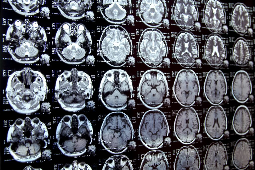
Medical image segmentation is a critical step in clinical research, providing insights into various aspects of patient health. This process involves annotating regions of interest in biomedical images, which can be time-consuming and labor-intensive, especially for complex structures like the brain’s hippocampus. Researchers at MIT have developed an AI-driven solution, MultiverSeg, that significantly streamlines this process by allowing users to interact with images in a more intuitive way.
Understanding the Importance of Segmentation
In clinical studies, analyzing how organs or structures change over time often requires precise delineation of those areas in imaging scans. For instance, studying how the hippocampus alters with age involves circling the hippocampus in multiple scans. Traditionally, researchers manually perform this labor-intensive task, which can limit the number of images they can process in a day. Such inefficiencies often hinder scientific exploration and delay advancements in medical treatments.
Introducing MultiverSeg: An AI-Powered Solution
Recognizing these challenges, MIT researchers have created MultiverSeg, a groundbreaking system that simplifies the segmentation of biomedical images. By leveraging user interactions—such as clicks, scribbles, and box drawings—MultiverSeg allows for rapid segmentation across entire datasets without requiring dedicated machine-learning expertise or extensive computational resources.
Key Features of MultiverSeg
-
User-Friendly Interaction: Clinicians can segment images easily through simple interactions. As the user annotates more images, the AI reduces the number of required interactions, ultimately reaching a point where no user input is needed for accurate segmentation.
-
Contextual Learning: Unlike traditional models that operate independently on each image, MultiverSeg utilizes context from previously annotated images. This architecture is designed to improve predictions, drawing from a context set of any size for guidance.
-
No Training Data Required: Users can begin a new segmentation task without needing a presegmented dataset, making it easy to utilize MultiverSeg for various medical imaging tasks with minimal setup.
-
Time Efficiency: As researchers using MultiverSeg continue to interact with the tool, it becomes progressively adept, drastically cutting down on time spent manually segmenting images.
-
High Accuracy: Performance evaluations have showcased MultiverSeg outperforming other state-of-the-art interactive segmentation systems, achieving 90% accuracy with 66% fewer scribbles and 75% fewer clicks.
Streamlining the Segmentation Process
Previously, researchers relied on either interactive segmentation tools or task-specific models. Interactive tools allowed for direct image input and marking but required repetition for each new image. Task-specific models necessitated extensive manual segmentation of hundreds of images to form a training dataset, followed by the training of a machine-learning model from scratch for new tasks.
MultiverSeg elegantly combines the strengths of both methods. It allows for intuitive interactions while maintaining a contextual reference that enhances accuracy with less user input. For instance, for particular image types like X-rays, users may only need to manually segment one or two images before the system is proficient enough to predict segmentations independently.
Enhancing Accuracy through Interactivity
One of the notable features of MultiverSeg is that it not only simplifies the segmentation process but also enables users to make corrections iteratively. This interactive capability allows researchers to refine the model’s predictions, enhancing accuracy until the desired results are achieved.
Hallee Wong, an electrical engineering and computer science graduate student and lead author of the research, emphasizes that “users can provide more interactions to refine AI predictions. This still dramatically accelerates the process because it is usually faster to correct an existing mistake than to start from scratch.”
Future Applications and Testing
The potential of MultiverSeg extends beyond enhancing the research landscape. It could play a pivotal role in improving clinical applications, such as radiation treatment planning. Streamlined segmentation could help reduce clinical trial costs and speed up the development of new treatment methods.
The research team envisions testing MultiverSeg in real-world scenarios alongside clinical collaborators, seeking further improvements based on user feedback. They also aim to enhance the system’s capabilities to accommodate the segmentation of 3D biomedical images in future iterations.
Conclusion
With MultiverSeg, MIT researchers have made significant strides in medical image segmentation, making the process quicker, more efficient, and accessible for researchers without extensive machine-learning backgrounds. This innovative tool has the potential to revolutionize how clinical studies are conducted, paving the way for faster scientific discoveries and improved patient care.
As the field of biomedical imaging continues to evolve, technologies like MultiverSeg will likely prove invaluable in speeding up research timelines and refining treatment methodologies. Supported by institutions such as Quanta Computer, Inc., and the National Institutes of Health, the ongoing development of this tool holds promise for a new era in medical research and patient outcomes.
The future of segmentation is here, and it could lead to breakthroughs in clinical practice, scientific understanding, and patient treatment plans.
Thanks for reading. Please let us know your thoughts and ideas in the comment section.
Source link
#system #accelerate #clinical #research #MIT #News